Research dossier
Retatrutide — GIP, GLP-1 and glucagon triple agonist dossier
An agonist at all three receptors — GIP, GLP-1 and glucagon — in clinical development.
Translated from the French original, which remains the reference version: lire en français →
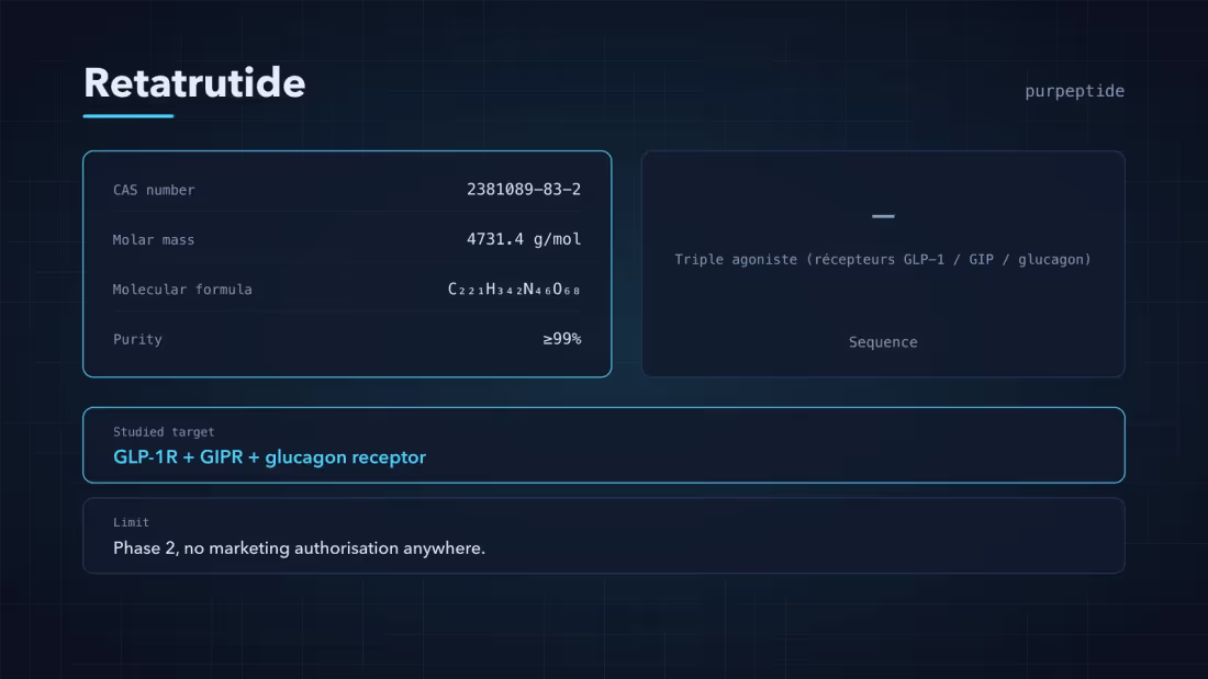
Molecular identity
| Parameter | Value |
|---|---|
| CAS number | 2381089-83-2 |
| Molar mass | 4731.4 g/mol |
| Molecular formula | C₂₂₁H₃₄₂N₄₆O₆₈ |
| Sequence / nature | Triple agonist (GLP-1 / GIP / Glucagon receptor) |
| Purity | ≥99% |
| Appearance | White lyophilised powder |
The values above correspond to the catalogue lot and are reproduced as they stand from the product page. Any discrepancy between this dossier and the product page should be reported before use.
Research context
Interest in this compound rests on a precise pharmacological question: does triple agonism deliver something that dual agonism does not? Answering it requires cell systems in which each receptor can be isolated, which is why recombinant single-expression lines are central to the literature. For an analytical laboratory, the value of the material lies less in the compound itself than in having a standard of known purity against which an assay method can be calibrated.
Mechanism studied
Retatrutide is described in the literature as a synthetic agonist acting at three G protein-coupled receptors of the secretin family: the GLP-1 (glucagon-like peptide-1) receptor, the GIP (glucose-dependent insulinotropic polypeptide) receptor and the glucagon receptor.
This triple affinity distinguishes it from the single agonists and from the GLP-1/GIP dual agonists described earlier. Published receptor pharmacology has measured binding affinity and adenylate cyclase activation for each of the three targets separately, in recombinant cell systems expressing one receptor at a time — a methodology that allows the respective contributions to be separated.
The downstream signalling described for these three receptors converges on the cAMP/PKA pathway, with β-arrestin recruitment profiles that differ by receptor and by ligand. That difference in recruitment — signalling bias — receives particular attention in the methodological literature, because it governs receptor internalisation and therefore the response kinetics observed in culture.
In-vitro literature
Published in-vitro characterisation concerns mainly competitive binding assays and intracellular cAMP measurement in cell lines expressing the recombinant human receptors. Phase III work on body weight has been carried out on this compound and is referenced in the public clinical literature; it falls outside the scope of use of this reference material and is mentioned here only as bibliographic context.
For analytical use, the relevant parameters are chromatographic purity, identity confirmation by mass spectrometry, and the actual peptide content per vial — that last value being the one that determines the effective concentration of a reconstituted solution.
Bibliographic entry points
- PubMed — peer-reviewed literature
- PubChem — chemical profile and assay data
- NCBI — methodology and receptor references
These links are an entry point into the literature, not an edited selection. None of the sources cited relates to a human use of this material, and consulting them directly is recommended for any protocol design.
Distinguishing this compound from its analogues
Not to be confused with the GLP-1/GIP dual agonists, nor with the single GLP-1 agonists, whose receptor profiles differ. The CAS number is the only reliable identifier: trade names and development codes vary from one source to another.
The CAS number shown on the product page is the identifier to rely on. Trade names, development codes and customary abbreviations vary from one source to another and are not a reliable identification.
Architecture moléculaire
Retatrutide is a synthetic peptide of high molecular mass — 4,731.4 g/mol — built on a glucagon-derived scaffold. That structural kinship is not incidental: glucagon, GLP-1 and GIP all belong to the secretin-glucagon superfamily and share a common general organisation, which is what makes it possible to design a single sequence able to engage all three receptors.
Three design elements distinguish the molecule.
The non-natural substitutions. Several positions carry non-proteinogenic amino acids, notably α-aminoisobutyric acid (Aib). These residues locally rigidify the helix and, above all, block cleavage by dipeptidyl peptidase-4 (DPP-4), the enzyme that inactivates endogenous GLP-1 within minutes.
The lipidation. A fatty acid chain is grafted onto the sequence through a spacer. That chain binds reversibly to serum albumin, which sequesters the peptide in circulation and very substantially slows its renal elimination. It is what permits weekly rather than daily administration in the published protocols.
The balance of the three activities. The relative potency at each of the three receptors is a tuned parameter, not a consequence. Glucagon agonism that was too pronounced would tend to raise blood glucose; the concomitant GLP-1/GIP agonism has to offset that effect. The molecule is a deliberate pharmacological compromise.
Pharmacology of the three receptors
All three targets are class B G protein-coupled receptors. Their simultaneous engagement by a single ligand is the notable feature of this molecule.
| Receptor | Main pathway | Effect described in the literature |
|---|---|---|
| GLP-1R | Gs → cAMP → PKA | Glucose-dependent insulin secretion; slowing of gastric emptying; central satiety signalling |
| GIPR | Gs → cAMP | Potentiation of the insulin response; effects on adipose tissue |
| GCGR | Gs → cAMP; hepatic glycogenolysis | Associated with an increase in energy expenditure |
The third row is what differentiates the compound. The first two targets act mainly on energy intake — less consumed, earlier satiety. Glucagon receptor agonism is associated in the literature with an action on expenditure. The designers’ working hypothesis is that acting on both sides of the energy balance at once produces an effect of a magnitude neither reaches alone.
That hypothesis is consistent with the published data without being demonstrated by it: a clinical trial measures an outcome, not the mechanism that produced it. The published clinical data are treated separately.
Impurity profile and analytical considerations
This is the point that directly concerns a laboratory receiving a lot.
A peptide of 4,731 g/mol carrying non-natural residues and a lipidation is made by solid-phase synthesis over a large number of coupling cycles. Every cycle is an opportunity for failure, and the resulting impurity families are predictable:
Truncated sequences (deletion sequences). A failed coupling produces a chain missing one residue. These species are structurally very close to the intended product and often co-elute with it in HPLC, which makes them hard to resolve.
Products of incomplete lipidation. The fatty acid chain may not be grafted at all, or may be grafted at an incorrect position. The mass then differs clearly — which mass spectrometry detects unambiguously.
Species from incomplete deprotection. Residual protecting groups left in place after cleavage.
This profile is why, for this compound in particular, mass spectrometry is not a decorative supplement to the chromatogram. A high HPLC purity establishes that one component dominates the detected area; only the measured mass establishes that this component really is the 4,731.4 g/mol molecule and not a truncated sequence resembling it. The guide to reading an analysis report sets out that cross-reading.
Stability and degradation
Lyophilised and held at −20 °C away from light, the compound is stable on a scale of months. In solution, several degradation routes become active.
Hydrolysis of the peptide bonds, accelerated in acidic or basic media and by temperature.
Deamidation of asparagine and glutamine residues, which changes the mass by +1 Da per event — a small difference, but detectable by high-resolution mass spectrometry.
Oxidation of susceptible residues, favoured by light and dissolved oxygen.
Aggregation, favoured by mechanical agitation. That is the operational reason a reconstituted vial should not be shaken: the air-liquid interface created by foaming denatures peptides.
Methodological limits
Triple agonism is precisely what complicates interpretation: an effect measured in a cell system expressing all three receptors cannot be attributed to any one of them without parallel single-expression assays. Publications reporting only multi-receptor systems therefore support no conclusion about the respective contribution of each pathway. The high molecular mass and the lipid chain also cause appreciable adsorption to plastic surfaces, a source of lost effective concentration that is rarely controlled.
These limits are not formalities: they determine what a protocol using this material can and cannot establish. Making them explicit in the experimental plan avoids attributing to the compound an observation that belongs to the measurement system.
Analytical parameters
The high molecular mass and the presence of a fatty acid chain in the structure make the chromatographic separation sensitive to the composition of the mobile phase. The RP-HPLC profile obtained with an acetonitrile/TFA gradient on a C18 column is the purity reference adopted.
| Parameter | Value |
|---|---|
| Storage | Store at −20 °C, protected from light |
| Solubility | Soluble in bacteriostatic water (BAC) |
| Stability | Stable for 24 months at −20 °C; 30 days after reconstitution at 2–8 °C |
| Control methods | RP-HPLC · MS · NMR |
Reconstitution is a matter for the experimental protocol. The volume of solvent determines the final concentration and is not prescribed here: it depends on the design of the experiment and the conditions of the assay.
Resulting concentrations
The table below is arithmetic: it gives the concentration obtained for a given solvent volume, with no recommendation of use.
| Format | 1 ml | 2 ml | 3 ml |
|---|---|---|---|
| 5mg | 5.00 mg/ml | 2.50 mg/ml | 1.67 mg/ml |
| 10mg | 10.00 mg/ml | 5.00 mg/ml | 3.33 mg/ml |
| 15mg | 15.00 mg/ml | 7.50 mg/ml | 5.00 mg/ml |
| 30mg | 30.00 mg/ml | 15.00 mg/ml | 10.00 mg/ml |
These values assume complete dissolution and a peptide content consistent with the lot report. Where the actual quantitative content differs from the nominal mass — a common discrepancy, caused by counter-ions and residual water — the effective concentration is lower than the theoretical value. That is precisely why quantitative content per vial forms part of the verification protocol.
Protocol design
Three points govern the validity of an assay using this material, whatever the question being asked.
The effective concentration is not the nominal concentration. It depends on the actual peptide content of the lot, on complete dissolution and on adsorption to the walls of the container. A concentration check after reconstitution is preferable to assuming a perfect yield.
The actual exposure time may be shorter than the nominal duration where the compound is degraded by the medium. Verify stability under the exact conditions of the assay — medium, serum, temperature, duration — before interpreting an absence of effect.
The control arm must correspond to the vehicle actually used, at the same final concentration. This requirement is unremarkable and remains the most frequent cause of irreproducible results.
Lot verification
The following formats are covered by a Janoshik Analytical report, readable from the product page:
- 10mg — lot report available
- 15mg — lot report available
- 30mg — lot report available
- 5mg — lot report available
| Format | Lot report | Availability |
|---|---|---|
| 5mg | Covered | In stock |
| 10mg | Covered | In stock |
| 15mg | Covered | In stock |
| 30mg | Covered | In stock |
The verification programme covers exclusively the identity and purity of the material, by reversed-phase chromatography and mass spectrometry. It covers neither sterility, nor endotoxins, nor any microbiological characteristic.
Frequently asked questions
How does this product arrive?
As a lyophilised powder in a sealed vial, shipped in insulated, plain packaging. Return the vial to the storage temperature given in the specifications on receipt.
Which solvent should be used for reconstitution?
Bacteriostatic water is the usual reconstitution solvent for lyophilised peptides in a laboratory setting. The volume is a matter for the experimental protocol and the concentration sought; we provide no dosage recommendation.
How long does the material remain stable?
The stability window appears in the specifications table on this page, before and after reconstitution. Those values correspond to the storage conditions stated; any temperature variation shortens them.
Does the analysis report correspond exactly to my vial?
The report linked on this page corresponds to the format selected. Each report is issued by Janoshik Analytical from an anonymously submitted sample, and covers the identity and purity of the peptide.
Can this product be used on humans or animals?
No. It is an analytical grade reference material intended exclusively for in-vitro laboratory research. Any administration to humans or animals is excluded, and no therapeutic, diagnostic or medical claim is made.
Dossiers in the same category
- Cagrilintide — CAS 1415456-99-3